2.1.2 External anatomy
The most prominent feature of bivalves is the two valves of the shell that may or may not be equal and may or may not completely enclose the inner soft parts. They have a variety of shapes and colours depending on species. The valves are composed mostly of calcium carbonate and have three layers; the inner or nacreous layer, the middle or prismatic layer that forms most of the shell, and the outer layer or periostacum, a brown leathery layer which is often missing through abrasion or weathering in older animals.

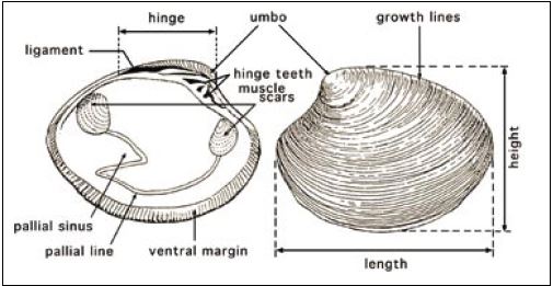
Figure 6: External and internal features of the shell valves of the hard shell clam, Mercenaria mercenaria. Modified from Cesari and Pellizzato, 1990.
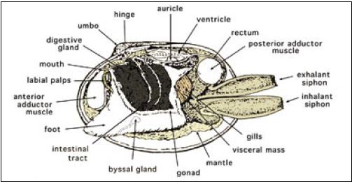
Bivalves do not have obvious head or tail regions, but anatomical terms used to describe these areas in other animals are applied to them. The umbo or hinge area, where the valves are joined together, is the dorsal part of the animal (Figure 6). The region opposite is the ventral margin. In species with obvious siphons (clams), the foot is in the anterior-ventral position and the siphons are in the posterior area (Figure 7). In oysters the anterior area is at the hinge and in scallops it is where the mouth and rudimentary foot are located.
There are right and left valves. Shell length is the greatest distance between the anterior and posterior margins of the shell; height is the greatest distance between the dorsal and ventral margins (Figure 6). The valves are joined at the umbo or hinge area by the hinge ligament and along this margin are irregular tooth-like projections called hinge teeth. These fit into grooves on the opposite valve and the interlocking arrangement fits the valves together. In species such as scallops a pit in the centre of the hinge is easily observed and it contains a dark elastic pad, the resilium. The hinge ligament and resilium spring the valves apart when the adductor muscle(s) relaxes.
The interior of the valves is smooth and shiny and may have a variety of colours depending on species. In species with anterior and posterior adductor muscles (i.e. clams and mussels) the two circular attachment scars are visible. In species with a single adductor muscle (monomyarian species) a single muscle scar is visible near the centre of the shell. About one third the distance in from the edge of the shell there is a shallow indistinct groove, the pallial line, that marks the attachment of the mantle.

Figure 7: The internal, soft tissue anatomy of a clam of the genus Tapes. In this view, the uppermost gill lamellae have been removed to reveal the foot and other adjacent tissues. Modified from Cesari and Pellizzato, 1990.