2.1.3 Internal anatomy
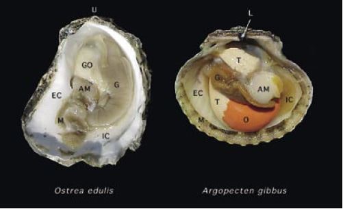
Careful removal of one of the shell valves reveals the soft parts of the animals. The differences in general appearance of an oyster and scallop can be seen in Figure 8.

Figure 8: The soft tissue anatomy of the European flat oyster, Ostrea edulis, and the calico scallop, Argopecten gibbus, visible following removal of one of the shell valves. Key: AM – adductor muscle; G – gills; GO – gonad (differentiated as O – ovary and T – testis in the calico scallop); L – ligament; M – mantle and U – umbo. The inhalant and exhalant chambers of the mantle cavity are identified as IC and EC respectively.
Mantle
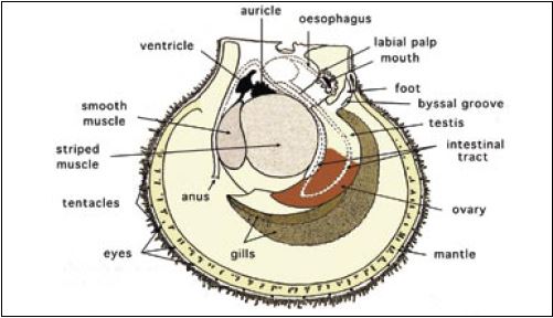
The soft parts are covered by the mantle, which is composed of two thin sheaths of tissue, thickened at the edges. The two halves of the mantle are attached to the shell from the hinge ventral to the pallial line but are free at their edges. The thickened edges may or may not be pigmented and have three folds. The mantle edge often has tentacles; in clams the tentacles are at the tips of the siphon. In species such as scallops the mantle edge not only has tentacles but also numerous light sensitive organs – eyes (Figure 9).

Figure 9: The internal, soft tissue anatomy of a hermaphroditic scallop.
The main function of the mantle is to secrete the shell but it also has other purposes. It has a sensory function and can initiate closure of the valves in response to unfavourable environmental conditions. It can control inflow of water into the body chamber and, in addition, it has a respiratory function. In species such as scallops, it controls water flow into and out of the body chamber and hence movement of the animal when it swims.
Adductor muscle(s)
Removal of the mantle shows the underlying soft body parts, a prominent feature of which are the adductor muscles in dimyarian species (clams and mussels) or the single muscle in monomyarian species (oysters and scallops). In clams and mussels the two adductor muscles are located near the anterior and posterior margins of the shell valves. The large, single muscle is centrally located in oysters and scallops. The muscle(s) close the valves and act in opposition to the ligament and resilium, which spring the valves open when the muscles relax. In monomyarian species the divisions of the adductor muscle are clearly seen. The large, anterior (striped) portion of the muscle is termed the “quick muscle” and contracts to close the valves shut; the smaller, smooth part, known as the “catch muscle,” holds the valves in position when they have been closed or partially closed. Some species that live buried in the substrate (e.g. clams) require external pressure to help keep the valves closed since the muscles weaken and the valves open if clams are kept out of a substrate in a tank.
Gills
The prominent gills or ctenidia are a major characteristic of lamellibranchs. They are large leaf-like organs that are used partly for respiration and partly for filtering food from the water. Two pairs of gills are located on each side of the body. At the anterior end, two pairs of flaps, termed labial palps, surround the mouth and direct food into the mouth.
Foot
At the base of the visceral mass is the foot. In species such as clams it is a well developed organ that is used to burrow into the substrate and anchor the animal in position. In scallops and mussels it is much reduced and may have little function in adults but in the larval and juvenile stages it is important and is used for locomotion. In oysters it is vestigial. Mid-way along the foot is the opening from the byssal gland through which the animal secretes a thread-like, elastic substance called “byssus” by which it can attach itself to a substrate. This is important in species such as mussels and some scallops enabling the animal to anchor itself in position.
Digestive system
The large gills filter food from the water and direct it to the labial palps, which surround the mouth. Food is sorted and passed into the mouth. Bivalves have the ability to select food filtered from the water. Boluses of food, bound with mucous, that are passed to the mouth are sometimes rejected by the palps and discarded from the animal as what is termed “pseudofaeces”. A short oesophagus leads from the mouth to the stomach, which is a hollow, chambered sac with several openings. The stomach is completely surrounded by the digestive diverticulum (gland), a dark mass of tissue that is frequently called the “liver”. An opening from the stomach leads to the much-curled intestine that extends into the foot in clams and into the gonad in scallops, ending in the rectum and eventually the anus. Another opening from the stomach leads to a closed, sac-like tube containing the crystalline style. The style is a clear, gelatinous rod that can be up to 8 cm in length in some species. It is round at one end and pointed at the other. The round end impinges on the gastric shield in the stomach. It is believed it assists in mixing food in the stomach and releases enzymes that assist in digestion. The style is composed of layers of mucoproteins, which release digestive enzymes to convert starch into digestible sugars. If bivalves are held out of water for a few hours the crystalline style becomes much reduced and may disappear but it is reconstituted quickly when the animal is replaced in water.
Circulatory system
Bivalves have a simple circulatory system, which is rather difficult to trace. The heart lies in a transparent sac, the pericardium, close to the adductor muscle in monomyarian species. It consists of two irregular shaped auricles and a ventricle. Anterior and posterior aorta lead from the ventricle and carry blood to all parts of the body. The venous system is a vague series of thin-walled sinuses through which blood returns to the heart.
Nervous system
The nervous system is difficult to observe without special preparation. Essentially it consists of three pairs of ganglia with connectives (cerebral, pedal and visceral ganglia).
Urogenital system
Sexes of bivalves can be separate (dioecious) or hermaphroditic (monoecious). The gonad may be a conspicuous, well defined organ as in scallops or occupy a major portion of the visceral mass as in clams. The gonad is generally only evident during the breeding season in oysters when it may form up to 50% of the body volume. In some species such as scallops, the sexes can be readily distinguished by eye when the gonad is full since the male gonad is white in colour and the female is red, even in hermaphroditic species. Colour of the full gonad may distinguish the sexes in some species such as mussels. In other species, microscopic examination of the gonad is required to determine the sex of the animal. A small degree of hermaphrodism may occur in dioecious species.
Protandry and sex reversal may occur in bivalves. In some species there is a preponderance of males in smaller animals indicating that either males develop sexually before females or that some animals develop as males first and then change to females as they become larger. In some species, e.g. the European flat oyster, Ostrea edulis, the animal may spawn originally as a male in a season, refill the gonad with eggs and spawn a second time during the season as a female.
The renal system is difficult to observe in some bivalves but is evident in such species as scallops where the two kidneys are two small, brown, sac-like bodies that lie flattened against the anterior part of the adductor muscle. The kidneys empty through large slits into the mantle chamber. In scallops, eggs and sperm from the gonads are extruded through ducts into the lumen of the kidney and then into the mantle chamber.