4.2.5 Fertilization procedures
Before fertilization, if not already done, egg suspensions should be gently filtered through a suitable mesh-size sieve (90 ?m aperture or greater) held so that the mesh is below water level in a larger volume bucket or container. This step is to remove contaminating faecal pellets from the adults prior to the addition of the sperm to reduce risk of the subsequent proliferation of bacteria and other micro-organisms during the next stage in the culture process.

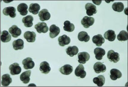
Figure 48: Dividing Crassostrea gigas eggs about 50 minutes after fertilization. Most of these eggs are developing normally and are at the 2 and 4-cell stage.
The method used to fertilize eggs is essentially the same whether for monoecious or dioecious species. The one exception in hermaphroditic bivalves is to ensure that eggs are cross-fertilized with sperm from adults other than the one that provided the particular batch of eggs. For this reason, batches of eggs from the different adults are kept separate and are separately fertilized with recently shed sperm from 3 or 4 males in the ratio 2 ml of sperm per l of egg suspension. Following sperm addition, they are allowed to stand for 60 to 90 minutes before pooling – if required – with the fertilized eggs from other adults.

Figure 49: Stages in the early development of eggs; A – sperm swarming around a rounded-off egg; B – extrusion of the first polar body following fertilization; C – two-cell stage also showing the second polar body; D – four-cell stage; E – eight-cell stage. The eggs of most oviparous bivalves range in size from about 60 to 80 ?m, depending on species. The time from fertilization to the various developmental stages is species and temperature dependent.
Within this time period, at the appropriate temperature for the species, the fertilized eggs will begin to divide, first almost equally into two cells and then unequally into 4 cells where one large cell will be observed capped by 3 much smaller cells. The first sign of successful fertilization, however, before cell division starts, is the extrusion from the egg of a small, transparent, dome-like structure, which is the first polar body (Figures 48 and 49). Assessment of the percentage of eggs developing normally can be made using a relatively low power microscope (x20-40 magnification). Fertilization rates almost invariably exceed 90% assuming the eggs are fully mature.
80 Hatchery culture of bivalves. A practical manual
It is desirable to estimate egg numbers prior to or within 20 to 30 minutes of fertilization since development will be impaired if the density of embryos per unit volume beyond the early stages of cleavage exceeds certain specified limits. This density is specified later and the method to determine both egg and larval numbers is described in section 5.1.2.3.